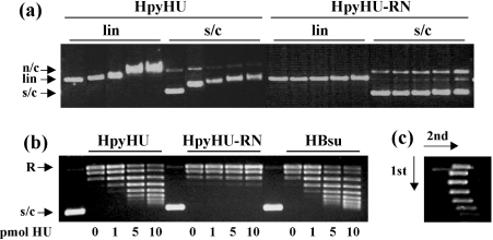
Figure 4. Interaction of HpyHU with plasmid DNA.
(a) Agarose gel electrophoresis of HpyHU variants binding to supercoiled (s/c) or linear (lin) DNA. n/c, nicked circular DNA. HpyHU variants are identified at the top, and protein concentrations, identical for all panels, are 0, 50, 100, 200 and 250 nM from left to right. (b) HpyHU introduces DNA supercoiling. Plasmid relaxed with topoisomerase I is supercoiled in the presence of HpyHU (left panel) or HBsu (right panel). HpyHU-RN is nearly inactive (middle panel). Protein concentrations are indicated below the panels. Relaxed (R) and supercoiled (s/c) DNA is indicated on the left. Lanes 1, 6 and 11 (numbered from the left) contain plasmid DNA not treated with topoisomerase I. (c) Two-dimensional electrophoresis of DNA topoisomers generated in the presence of 10 pmol of HpyHU. Negatively supercoiled DNA loses superhelicity in the presence of chloroquine, forming the left branch of the arc. The directions of the first and second dimensions are indicated by arrows.

