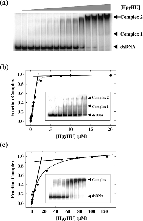
Figure 5. Determination of the non-specific site size for HpyHU.
(a) Titration of HpyHU with 1.0 μM perfect duplex 37 bp DNA [25]. Complex and free DNA is identified at the right. [HpyHU], 0–20 μM. (b) Binding isotherm corresponding to the titration shown in (a). The breakpoint corresponds to the molar equivalence between ligand concentration and lattice residues, defining the occluded non-specific site size. The inset shows formation of two distinct complexes with increasing concentration of HpyHU binding to 37 bp duplex at 10 mM KCl. Protein concentrations are 0, 5, 10, 25, 50, 100, 250 and 500 nM from left to right respectively. (c) Binding isotherm for HpyHU binding to 3.5 μM 80 bp DNA. Inset shows representative titration of HpyHU with 80 bp DNA.

