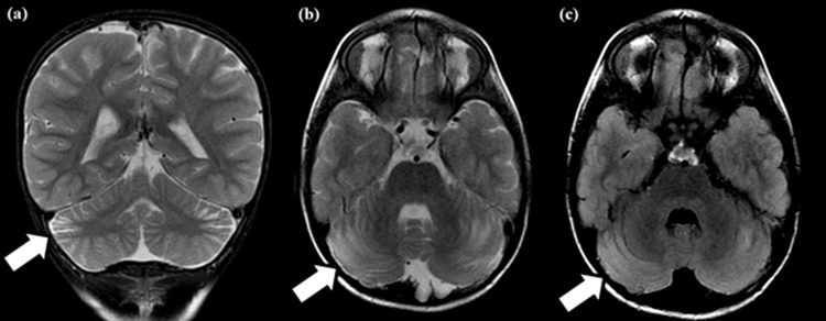
Figure 2. Coronal T2-weighted (a), axial T2-weighted (b), and fluid-attenuated inversion recovery (c) MRIs.
Cortico-subcortical bands with hyperintensities are noted in the periphery of the middle third of the cerebellar hemispheres, associated with slight regional volumetric reduction (white arrows). These findings likely indicate regional gliosis with slight atrophy.

