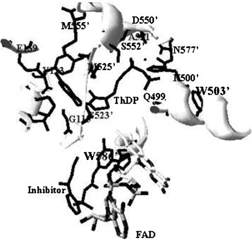
Figure 7. Structural representation of the ThDP-binding site.
The active site centred at ThDP was modelled by a SwissProt database program using the X-ray co-ordinates of yeast AHAS (Protein Data Bank code 1N0H). The three-dimensional structure represented here was constructed with the inhibitor, chlorimuron ethyl. Trp586′ was shown only when inhibitor was added to the crystal formation. Trp503′ and Trp586′ correspond to residues Trp490 and Trp573 of tobacco AHAS respectively. Protein side chains close to ThDP or within 5 Å to the likely position of active sites are shown, with residues from the domain of one subunit designated as a number and those from the other subunit as a primed number.

