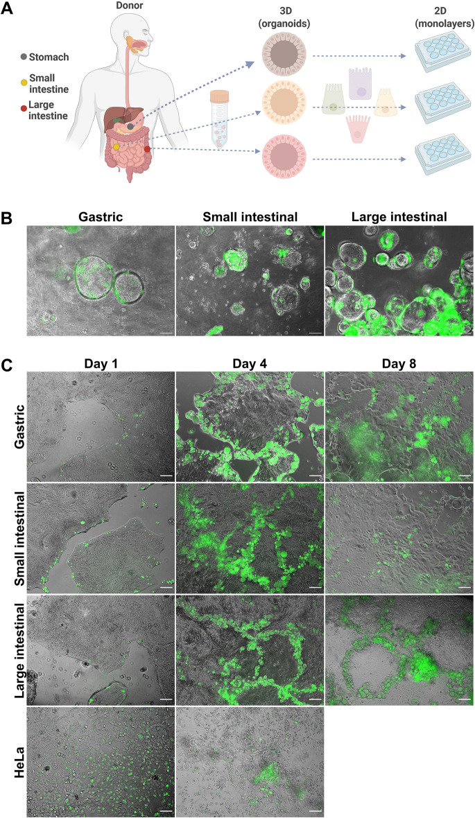
Fig 1. C. trachomatis infects patient-derived GI cells.
(A) Schematic representation of host model generation. (B) Human gastric and intestinal organoids infected with GFP-expressing C. trachomatis (MOI of 5) at 48 hours p.i. (C) Organoid-derived subconfluent monolayers of human primary gastrointestinal epithelial cells and HeLa cells were infected with GFP-expressing C. trachomatis (MOI of 0.5) and observed daily. Shown are representative images obtained at 1, 4 and 8 days p.i.. Images in (B) and (C) were taken with phase-contrast and in the green fluorescence channel and merged. Scale bar: 100 μm. Fig 1A was prepared in Biorender.com.

