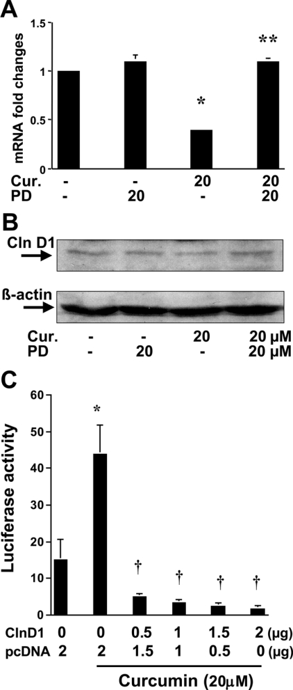
Figure 1. Activation of PPARγ mediates curcumin suppression of cyclin D1 expression in passaged HSC, which, in turn, facilitates the activation of PPARγ.
Passaged HSC were pretreated with or without the PPARγ antagonist PD 68235 (20 μM) for 30 min prior to the addition of curcumin (Cur.) at 20 μM for an additional 24 h. Total RNA and protein extracts were prepared for real-time PCR (A) (n=3) and Western blotting analyses (B) (n=3) respectively. Significance: *P<0.05 compared with cells without curcumin; **P<0.05 compared with cells with curcumin. (C) To assess the effects of cyclin D1 (ClnD1) on PPARγ activity, HSC in 6-well culture plates were co-transfected with a total of 4.5 μg of plasmid DNA per well, including 2 μg of pPPRE-TK-Luc, 0.5 μg of pSV-β gal, pCMV-cyclinD1 at the indicated doses and an empty vector. The amount of DNA in pCMV-cyclinD1 plus the empty vector was 2 μg. After recovery, cells were treated with or without curcumin at 20 μM for 36 h. Luciferase activities were expressed as relative units after β-galactosidase normalization (n=6). Significance: *P<0.05 compared with cells without curcumin; †P<0.05 compared with cells transfected with no pCMV-cyclinD1, with curcumin treatment (second column).

