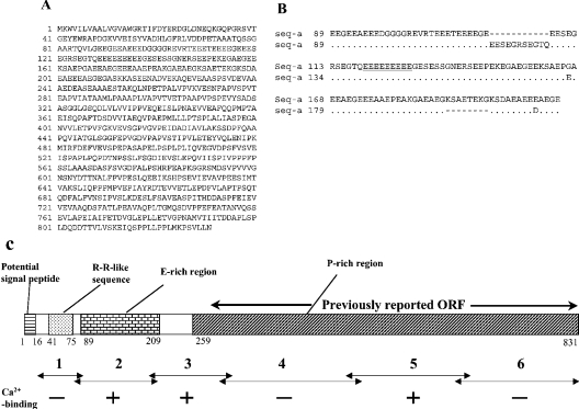
Figure 2. Primary structure of the encoded protein.
(A) Translated amino acid sequence of seq-a. Positions of amino acid residues are indicated on the left. (B) Alignment of the conceptual amino acid sequences of seq-a and b in the E-rich region. A dot and a hyphen indicate an identical amino acid and a gap respectively. The nine consecutive E residues are underlined. (C) A schematic drawing of the primary structure of CCN. Numbers below the boxes indicate positions of amino acid residues (in seq-a) at the boundaries of regions. Ca2+-binding (see text and Figure 4) was detected in the three fragments marked with +, but not in those marked with − (see Figure 5 and [24]).

