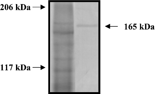
Figure 3. Western blot analysis of CCN.
An extract of an intermoult exoskeleton was subjected to SDS/PAGE, and proteins were detected with Coomassie Brilliant Blue (left-hand lane). Positions of molecular-mass standards are indicated on the left. In a Western blot (right-hand lane), an immunoreactive band is observed at 165 kDa (arrow).

