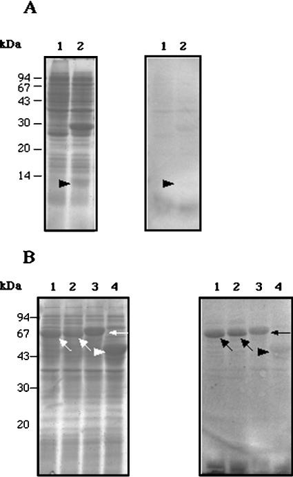
Figure 5. Ca2+ binding to bacterially expressed CCN fragments.
(A) Left-hand gel: SDS/PAGE of extracts of the following E. coli cells: lane 1, extracts of E. coli without an expression construct (a negative control); lane 2, extracts of E. coli expressing Fr1. Right-hand gel: 45Ca2+ overlay of a Western blot. Significant binding of Ca2+ to Fr1 is not detected (arrowhead). (B) Left-hand gel: SDS/PAGE of extracts of E. coli expressing the following proteins: lane 1, MBP–Fr2a; lane 2, MBP–Fr2b; lane 3, MBP–Fr3, lane 4, MBP–lacZα (negative control). The three fragment fusion proteins and MBP–lacZα are indicated with arrows and an arrowhead respectively. Right-hand gel: 45Ca2+ overlay of a Western blot. Binding of Ca2+ to the three fragment fusion proteins is detected in lanes 1, 2 and 3 (arrows), whereas no significant Ca2+-binding to MBP–lacZα is observed (arrowhead, lane 4).

