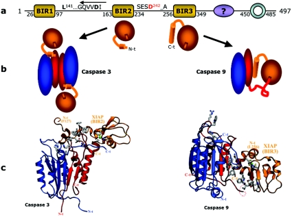
Figure 10. Mechanisms of caspase inhibition by XIAP.
(a) Schematic diagram of the domain organization of human XIAP. The sequence critical for the inhibition of effector caspases by XIAP is given [254,256]; the arrow above the sequence indicates that it binds in a ‘reverse mode’ to effector caspases (see text and Figure 11b). A caspase cleavage site in XIAP [253,263] is indicated; other cleavages are also detected [166]. (b) Cartoon depicting the mode of binding of BIR2 to caspase-3 and of BIR3 to caspase-9 (caspase large subunits in blue, and small subunits in red) bound to the respective BIR (orange). The shallow groove on each BIR domain symbolizes the Smac pocket into which the four N-terminal residues constituting an IAP-binding motif fit. Although important for caspase-9 inhibition [122], the significance of this groove in BIR2 is less well understood in terms of caspase-3 or -7 inhibition [255]. (c) Ribbon diagram of the inhibitory interactions, with the same colour coding. The N- and C-termini of the subunits of the complex are labelled, and important side chains in the interactions are shown, but for simplicity are not labelled.

