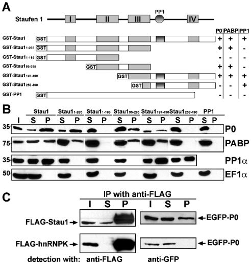
Figure 5. Interaction of Stau1 with ribosomal protein P0.
(A) Schematic representation of Stau1, showing its dRBDs I–IV (boxes) and PP1 interaction domain (circle), as well as different GST fusion proteins. Table 1 summarizes whether the corresponding GST fusion proteins are able to pull-down rat brain P0, PP1 and PABP. (B) Western blots of GST pull-down experiments probed with specific antibodies against P0, PABP, PP1α and EF1α. Pull-downs were performed with the indicated GST fusion proteins and an adult rat brain lysate. P0 and PABP are only pulled-down by fusion proteins containing dsRBD III. PP1 precipitation requires the previously characterized PP1 interaction site. (C) Western blots of anti-FLAG immunoprecipitates obtained from HEK-293 cells co-expressing full-length or truncated versions of EGFP-tagged P0, together with either FLAG–Stau1 (upper row) or FLAG–hnRNPK (lower row). Recombinant proteins were detected with either anti-FLAG or anti-GFP antibodies. P0 co-immunoprecipitates with recombinant Stau1, but not with hnRNPK. I, input; S, supernatant fraction; P, pellet fraction.

