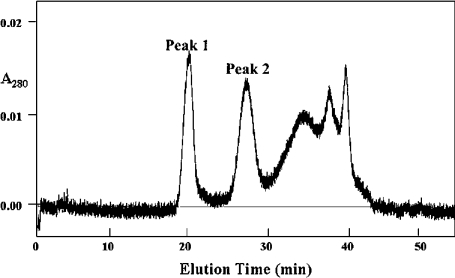
Figure 1. Separation of LPL1 (peak 1) and PLB1 (peak 2) from C. gattii by Superose 12 HR 10/30 column chromatography.
Freeze-dried active fractions post-UNO™Q-1 chromatography [8] were reconstituted in 50 mM Mes/2 mM EDTA/0.6 M NaCl, pH 6.3, and applied to the Superose column equilibrated in the same buffer. Proteins were eluted at a flow rate of 0.5 ml/min, and fractions were collected and assayed for LPL, LPTA and PLB activities.

