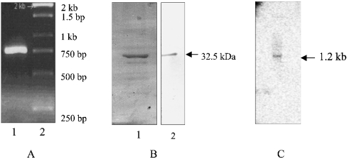
Figure 1. Preparation of PfΔFC.
RNA from the parasite was subjected to reverse transcriptase–PCR amplification with FC-specific primers, and the full-length cDNA obtained was sequenced. (A) Lanes: 1, PCR-amplified 750 bp fragment; 2, standards (1 kb ladder). (B) SDS/PAGE analysis of the C-terminal 32.5 kDa protein fragment expressed in E. coli and purified on an Ni2+-nitrilotriacetate column: Lanes: 1, Coomassie stain; 2, Western-blot analysis with anti-histidine tag antibodies. (C) Northern-blot analysis of parasite RNA with labelled PfΔFC cDNA showing an RNA band at 1.2 kb.

