Fig. 2.
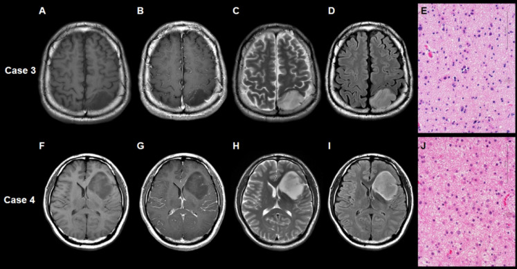
Case 3 is a representative case with left occipital lobe astrocytoma, IDH-mutant displaying the negative super T2-FLAIR mismatch sign and negative conventional T2-FLAIR mismatch sign. No evident contrast enhancement is observed (A, B). The T2-weighted (C) and FLAIR (D) images illustrate a tumor displaying a uniform hyperintensity. Hematoxylin-eosin staining showed (E) pleomorphic astrocyte tumor cells with a microcystic background. Case 4 is the representative case with left frontal lobe astrocytoma, IDH-mutant displaying a negative super T2-FLAIR mismatch sign but a positive conventional T2-FLAIR mismatch sign. No observable contrast enhancement was noted (F, G). The tumor manifests as a hyperintense area in the T2-weighted image (H) and exhibits a relatively low signal on the FLAIR image (I), except for a hyperintense peripheral rim. Hematoxylin-eosin staining (J) shows an astrocytic tumor amidst a fibrillary and microcystic background.

