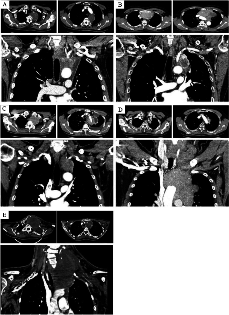
Fig. 2.
A. Computed tomography (CT) images showing a tumor with suspected invasion of the lung, aorta, vertebra and subclavian artery (Case 1). B. CT images showing a tumor with suspected invasion of the lung, hilar, and aorta (Case 2). C. CT images showing a tumor with suspected invasion of the lung, aorta, subclavian artery, and phrenic nerve (Case 3). D. CT images showing a tumor with suspected invasion of the lung, aorta, and subclavian artery, with possible extension to the neck (Case 4). E. CT images showing a tumor with suspected invasion of the left subclavian artery and subclavian vein, with possible extension to the neck (Case 5). SCA subclavian artery, SCV subclavian vein

