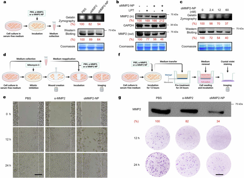
Fig. 4. Degradation of MMP2 mediated by αMMP2-NP.
a Gelatin zymography and Western blot assay of cell culture media for MMP2 activity and content. Media were collected after B16F10 cells were treated with PBS, αMMP2 (12 nM), or αMMP2-NP (α-MMP2-equiv. 12 nM) for 12 h. The gels and blot are representative of n = 3 biological replicates. b Western blot assay of MMP2 inside (IN) or outside (OUT) of B16F10 cells treated with αMMP2-NP (α-MMP2-equiv. 12 nM) for 12 h in the presence or absence of 0.1 mg mL−1 LPT. The gel and blots are representative of n = 3 biological replicates. c MMP2 activity and content in the culture media of B16F10 cells treated with varying concentrations of αMMP2-NP for 12 h. The gels and blot are representative of n = 3 biological replicates. d, e Schematic illustration (d) and results (e) of the wound-healing assay. B16F10 cells were treated with PBS, αMMP2 (12 nM), or αMMP2-NP (α-MMP2-equiv. 12 nM) for 12 h. Scale bar, 200 μm. The images are representative of n = 3 biological replicates. f, g Schematic illustration (f) and results (g) of the transwell cell invasion assay. CT26 cells were treated with PBS, αMMP2 (12 nM), or αMMP2-NP (α-MMP2-equiv. 12 nM). Scale bar, 200 μm. These images are representative of n = 3 biological replicates. All the uncropped gels and blots are included in the Source Data file. Source data are provided as a Source Data file.

