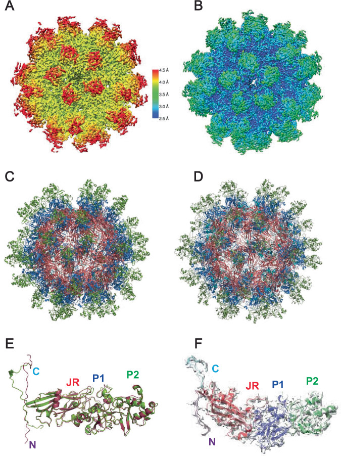
Fig. 3. Cryo-EM structures of ANV virus-like particles produced in insect and mammalian cells.
A The cryo-EM map demonstrating the local resolution of the SF9 cell expression purified LY1 ΔARM ORF1 particle colored by its resolution (shown on the scale to the right). B. The cryo-EM map marking the local resolution of expi293-expression system purified LY1 ΔC-Term particle colored by its resolution (same scale as in A). C A ribbon representation of the LY1 ΔARM 60-mer particle atomic model. D A ribbon representation of the LY1 ΔC-Term atomic model shown as in (C). E An overlay of the SF9 cell expression purified (red) and mammalian cell-derived (green) protomers. The observable N- and C-termini and jelly roll, P1 and P2 domains are labeled. F One ΔC-Term ORF1 protomer shown in its electron density with domains labeled and colored as above.

