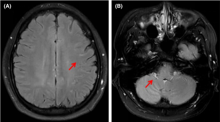
FIGURE 1.

(A) Contrast‐enhanced T2‐weighted magnetic resonance imaging (T2WI) shows that leptomeningeal appears as enhancing curvilinear segments following the gyral convolutions of the bilateral cerebral hemispheres (arrow). (B) Contrast‐enhanced T2WI FLAIR showing the significant enhancement of the bilateral cerebellar leptomeningeal (arrow).
