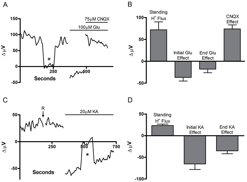
Figure 1.

Responses of cells with standing initial positive (acidic) H+ signals in response to application of glutamate and kainate measured using self-referencing H+ selective microelectrodes. (A) Representative trace showing the alkalinization response to application of 1mL bolus of 100 μM glutamate from one horizontal cell. The alkalization was reversed with additional application of 1mL of a solution containing glutamate (100 μM) and the ionotropic glutamate receptor blocker CNQX (75 μM). Asterisk in this and other figures shows control recording 200 μm away from the cell. (B) Average result from 6 cells before glutamate, initially after glutamate, 100 sec after glutamate, and following co-application of CNQX. (C) Representative trace from a different horizontal cell. 1mL bath application of additional Ringer’s solution (R) by itself did not change the standing differential recording. Addition of 1mL of kainate induced an extracellular alkalization. (D) Average result from 8 cells before kainate, initially after kainate, and 100s after kainate.
