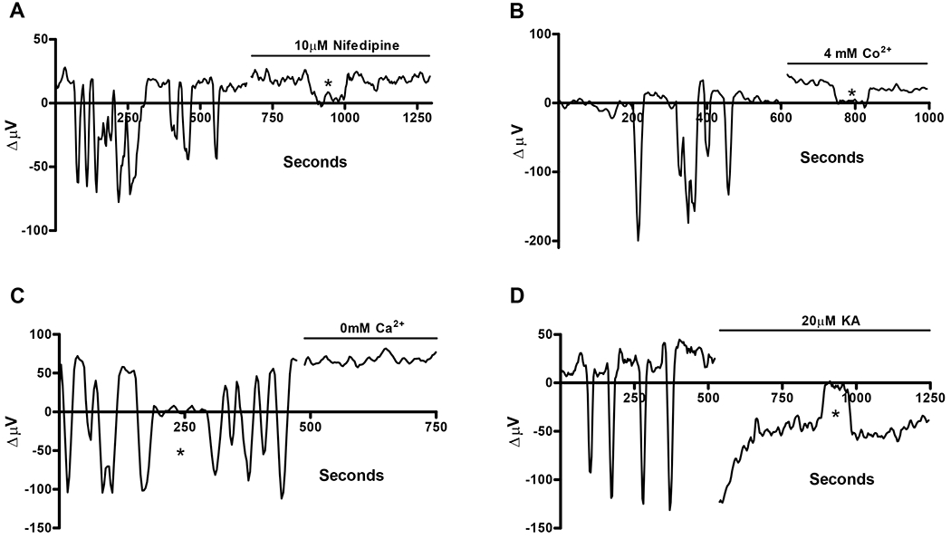
Figure 3.

Spontaneous extracellular H+ oscillations and their abolishment by nifedipine, cobalt, nominally 0mM extracellular Ca2+, and kainate. (A) Representative trace from one cell showing spontaneous extracellular H+ oscillations; addition of 10 μM nifedipine abolished the oscillations and resulted in a standing acidic H+ signal. (B) Representative trace showing oscillations from a second cell that were abolished reversed upon addition of 4 mM Co2+; a standing extracellular acidification was observed following cobalt application. (C) H+ oscillations observed in a third cell were eliminated by applying a solution containing nominally 0 mM external Ca2+. A continuous extracellular acidification was observed following the addition of the nominally 0 mM external calcium solution. (D) H+ oscillations in the final cell were quieted in an extracellular alkalinized state following the addition of 20 μM kainate.
