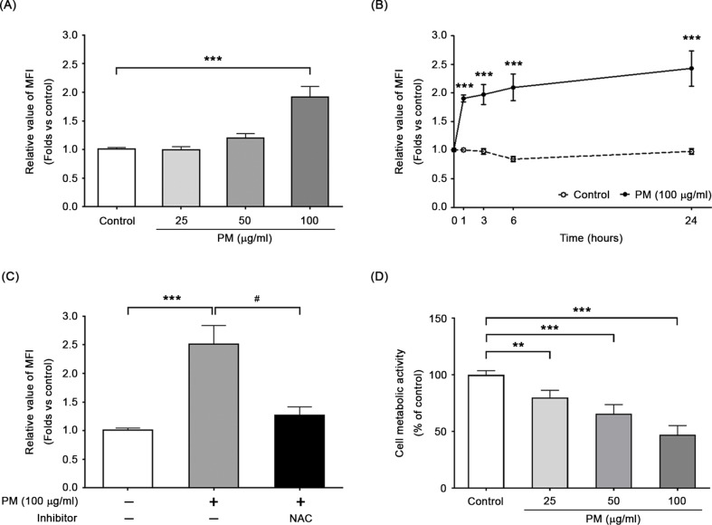
Fig. 1.
PM induced intracellular ROS formation in BEAS-2B cells. Intracellular ROS formation was detected using DCF-DA dye. The concentration (A) and time (B) dependent increases in the levels of intracellular ROS were observed. (C) ROS scavenger, NAC, significantly inhibited PM-induced ROS production in BEAS-2B cells. (D) In addition, the WST-1 assay was employed to assess metabolic activity of cells following exposure to PM. ***P < 0.001 and **P < 0.01, compared with untreated cells (control); ##P < 0.01 and #P < 0.05, compared with the PM-stimulated group (n = 3)

