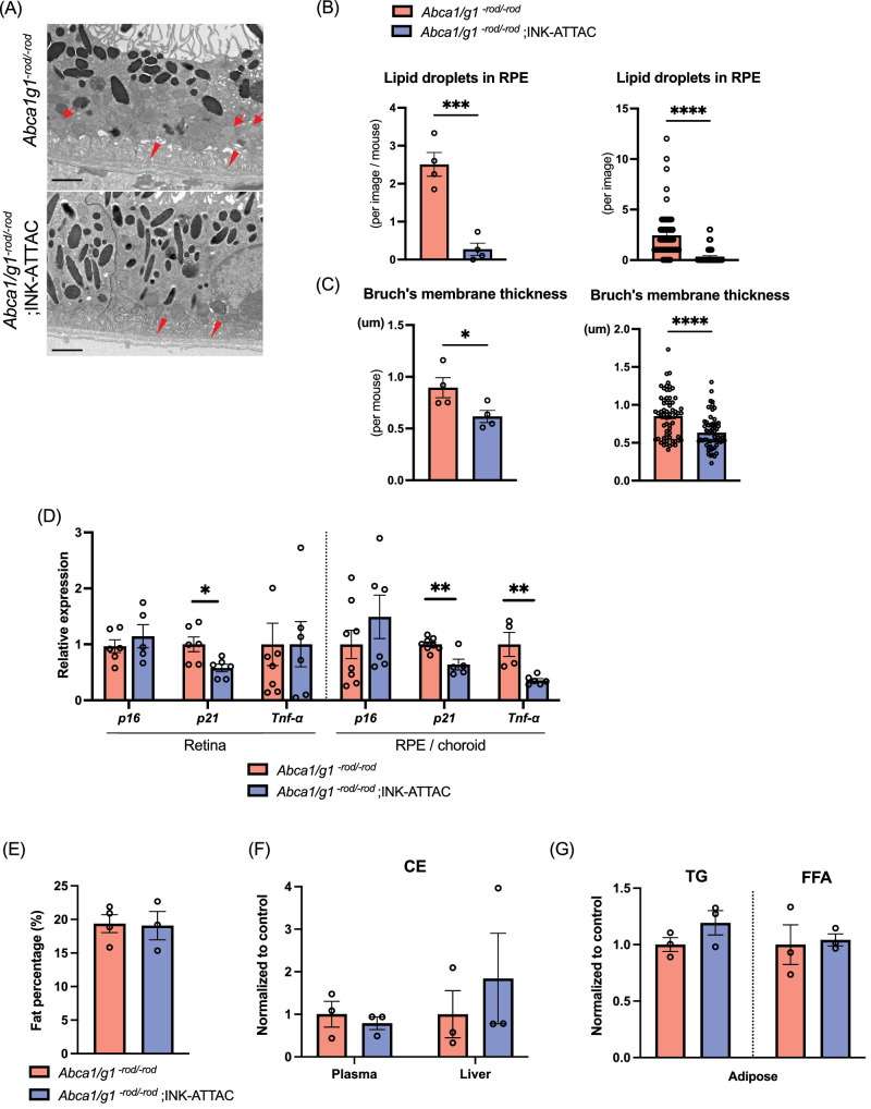
Figure 4.
Selective elimination of p16-positive senescent cells suppresses AMD-associated phenotype and senescence without altering systemic lipid metabolism. Representative electron microscopy images showing RPE cells and Bruch's membrane (A), and the quantification of the number of intracellular lipids in RPE cells (B) and Bruch's membrane thickness (C). Scale bar: 2 µm. Note intracellular lipids in RPE cells (arrow) and thickened Bruch's membrane (arrowhead) in Abca1/g1-rod/-rod. (D) mRNA expression of senescence markers (p16 and p21) and Tnf-α in the retina and RPE/choroid complex isolated from Abca1/g1-rod/-rod and Abca1/g1-rod/-rod;INK-ATTAC. Fat percentage (E), cholesterol ester in plasma and liver (F), and triglyceride and free fatty acids in adipose tissue (G) of Abca1/g1-rod/-rod and Abca1/g1-rod/-rod ;INK-ATTAC. *P < 0.05; **P < 0.01; ***P < 0.001; ****P < 0.0001, t-test. Data are represented as mean ± SEM.

