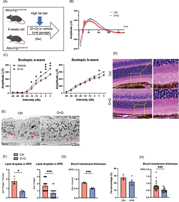
Figure 5.
Effects of pharmacological senolysis on retinal degeneration by cholesterol accumulation in photoreceptors. (A) Experimental design of senolytic drug administration (D+Q) for Abca1/g1-rod/-rod. (B) Electroretinography (ERG) waveform of Abca1/g1-rod/-rod treated with D+Q or vehicle. (C) The quantification of ERG (scotopic a-wave and b-wave) amplitudes. (D) Images of H&E staining. Scale bar: 50 µm. Representative electron microscopy images showing (E) RPE cells and Bruch's membrane, (F) the quantification of the number of intracellular lipids in RPE cells, and (G) Bruch's membrane thickness. Scale bar: 2 µm. Note that intracellular lipids in RPE cells (arrow) and thickened Bruch's membrane (arrowhead) in Abca1/g1-rod/-rod were treated with the vehicle. (H) Fat percentages of Abca1/g1-rod/-rod treated with D+Q. *P < 0.05; **P < 0.01; ***P < 0.001, t-test for comparison between two groups and two-way ANOVA followed by Bonferroni correction for comparison with multiple time points. Data are represented as mean ± SEM.

