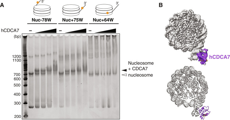
Fig. 3. Cryo-EM structure of hCDCA7 bound at linker DNA.
(A) EMSA analyzing the interaction of hCDCA7264–371 C339S with nucleosomes carrying hemimethylated CpG at the indicated positions. (B) A composite cryo-EM map (top) and the model structure (bottom) of hCDCA7264–371 C339S (generated from AF2) bound to Nuc+75W shown in (A). The map corresponding to CDCA7 is colored purple except for the conserved C-terminal helix, which is colored orange.

