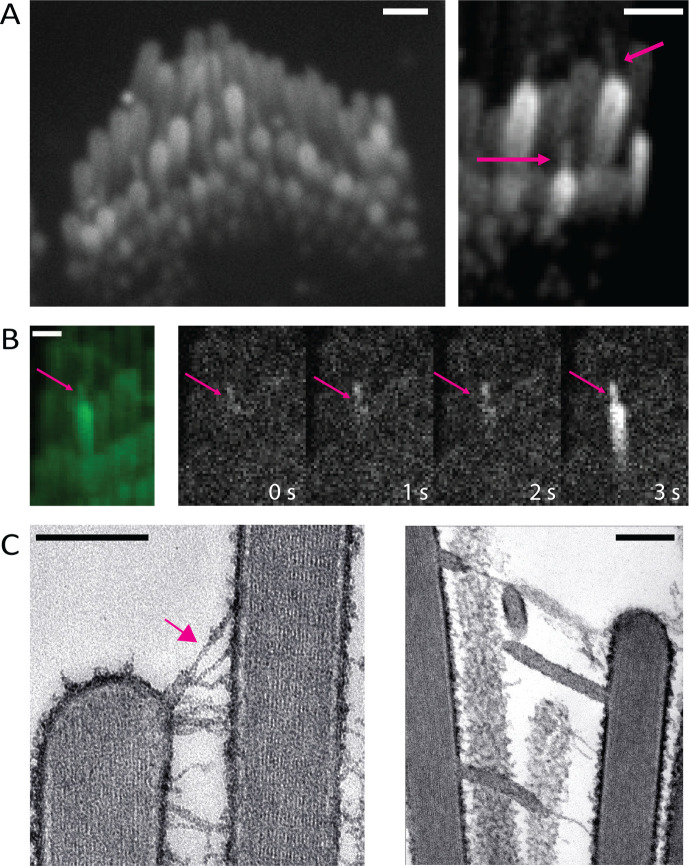
Figure 6: Tension in membrane tethers could play a role in generating calcium transients in stereocilia.
A) Over-deflected stereocilia experience a shear force against the coverslip. This results in the formation of membrane tethers which can be visualized with membrane-localized GCaMP3. The tethers can occur between two stereocilia or between the glass surface and a stereocilium (magenta arrows). A large proportion of the tethered stereocilia appear bright, indicating an increased calcium influx. Scale bars are1 μm. B) Membrane tethers play a role in calcium influx, and tethers can act as sites for the origination of spontaneous calcium transients. The intensity difference time series shown here was taken every 1s. It can be observed in this example that the membrane tether (magenta arrow) shows an increased calcium level first, followed by a calcium transient in the stereocilium. Scale bar is 1 μm. C) Over-deflected hair bundles in electron microscopy preparations display membrane tethers. The tip link remains intact at the tether end (magenta arrow, left). Scale bar is 200 nm.

