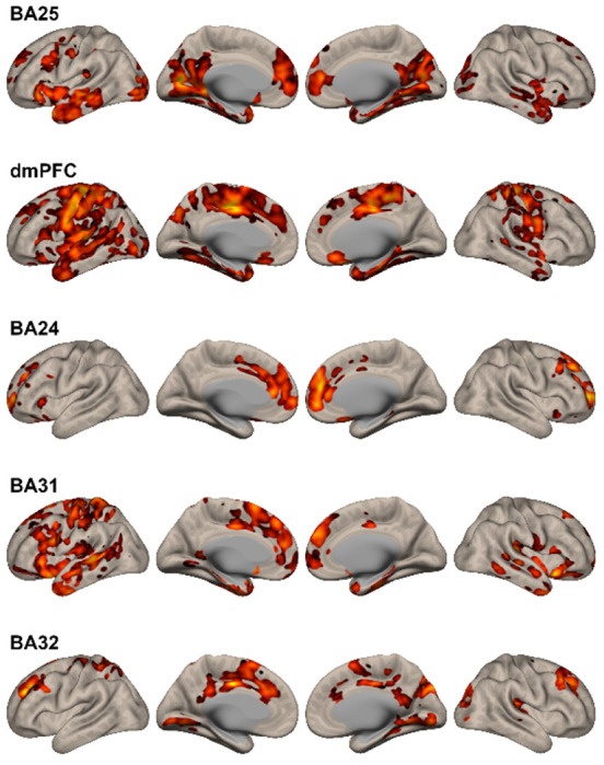
Figure 3. Changes in Functional Connectivity Observed After N2O Exposure.

Details of the regional changes observed for each seed are described in the text and Table 3S. In general, changes in functional connectivity in regions associated with mood regulation were observed, including the medial prefrontal cortex, anterior cingulate, paracingulate cortex, hippocampus, and parahippocampus. Additionally, all seeds demonstrated changes in functional connectivity in the dorsal paracingulate gyrus, previously identified as having increased resting-state connectivity in MDD as the “dorsal nexus” (Sheline et al., 2009).
