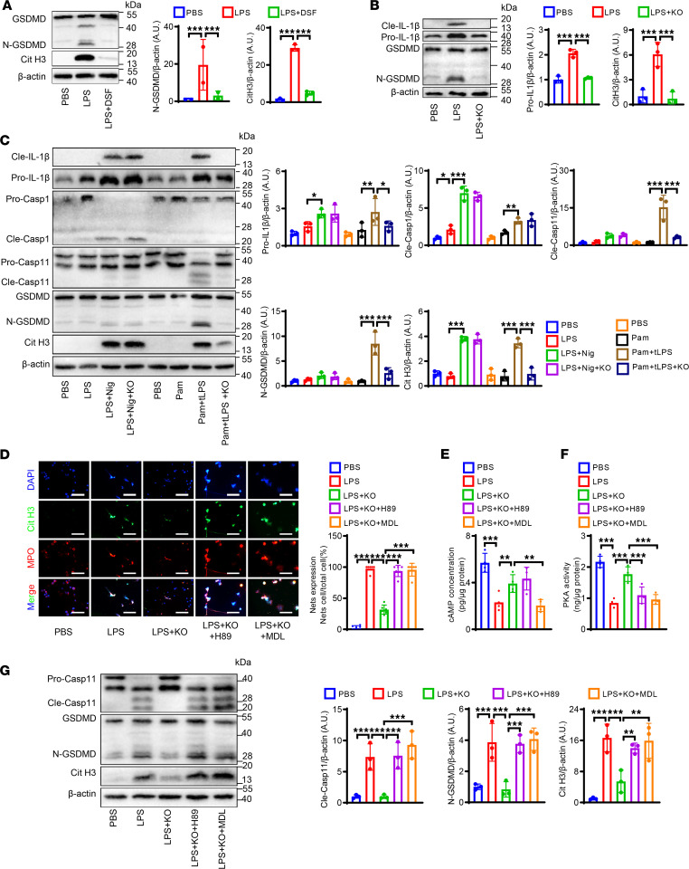
Figure 6. PI3Kγ promotes NETs’ formation via noncanonical pyroptosis pathways in vitro.
(A) Representative Western blot (A) and quantitative comparison of Cit H3 and GSDMD in WT neutrophils with different treatments: PBS, LPS (5 μg/mL), and LPS + DSF (30 μM). n = 3. (B) Representative Western blot and quantitative comparison of IL-1β and GSDMD in neutrophils with different treatments: PBS, LPS (5 μg/mL), LPS + KO (PI3Kγ–/–). n = 3. (C) Representative Western blot and quantitative comparison of Cit H3, IL-1β, GSDMD, caspase-11, and caspase-1 in the neutrophils with different treatments: PBS, LPS (100 ng/mL), LPS + nigericin (10 μM), LPS + nigericin + KO, PBS, Pam3CSK4 (1 μg/mL), Pam3CSK4 + transfer LPS (tLPS, 10 μg/mL), Pam3CSK4 + tLPS + KO. n = 3. (D) Representative immunofluorescence staining and quantitative comparison of NETs produced by neutrophils with different treatments: PBS, LPS (5 μg/mL), LPS + KO, LPS + H89 (20 μM) + KO, LPS + MDL12330A (10 μM) + KO. NETs were detected using immunofluorescence staining. NETs’ expression was calculated by NET-expressing cell numbers/total cell numbers per high-power field (original magnification, 40×). n = 6. Scale bars, 50 μm. (E) Comparison of cAMP concentration of neutrophils with different treatments as described in D by ELISA. n = 4. (F) Comparison of PKA kinase activity of neutrophils with different treatments as described in D by kinase activity assay. n = 4. (G) Representative Western blot and quantitative comparison of Cit H3, GSDMD, and caspase-11 in the neutrophils with different treatments as described in D. n = 3. (A–G) One-way ANOVA followed by Fisher’s least significant difference post hoc test. *P < 0.05, **P < 0.01, and ***P < 0.001. GSDMD, gasdermin D; pro-, prosoma; cle-, cleaved; Casp1, caspase-1; Casp11, caspase-11; LPS, lipopolysaccharide; DSF, disulfiram; Nig, nigericin; Pam, Pam3CSK4; KO, phosphoinositide-3-kinase γ knockout; MDL, MDL12330A.

