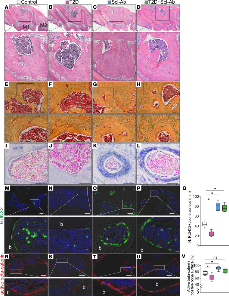
Figure 4. Scl-Ab treatment promotes robust bone formation.
(A–D) H&E staining shows the alveolar bone under the furcation area. (E–H) Pentachrome staining shows the bone around the marrow cavity. (I–L) ALP staining shows bone formation activity around the marrow cavity. (M–P) Immunostaining of RUNX2. (Q) Quantification of RUNX2+ bone-lining cells on the bone surface (n = 8). *P < 0.0001. (R–U) Immunostaining of active β-catenin. (V) Quantification of active β-catenin–positive bone surface (n = 8). *P < 0.001; ns, not significant. The data were analyzed using 1-way ANOVA with Tukey’s post hoc tests. Scale bars: 50 μm. b, bone; M1, maxillary first molar; M2, maxillary second molar.

