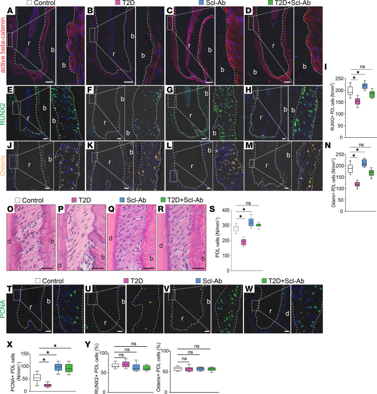
Figure 6. Scl-Ab treatment promotes PDL cell proliferation.
(A–D) Immunostaining of active β-catenin. (E–H) Immunostaining of RUNX2. (I) Quantification of RUNX2+ cells in the PDL (n = 8). *P < 0.05; ns, not significant. (J–M) Immunostaining of osterix. (N) Quantification of osterix+ cells in the PDL (n = 8). *P < 0.05. (O–R) H&E staining shows the PDL. (S) Quantification of PDL cells (n = 8). *P < 0.0001. (T–W) Immunostaining of PCNA. (X) Quantification of PCNA+ cells (n = 8). *P < 0.01. (Y and Z) Quantification of the percentages of (Y) RUNX2+ and (Z) osterix+ PDL cells (n = 8). Dotted lines indicate the demarcation between the PDL and alveolar bone or dentin/cementum. The data were analyzed using 1-way ANOVA with Tukey’s post hoc tests. Scale bar: 50 μm. r, roots; b, bone.

