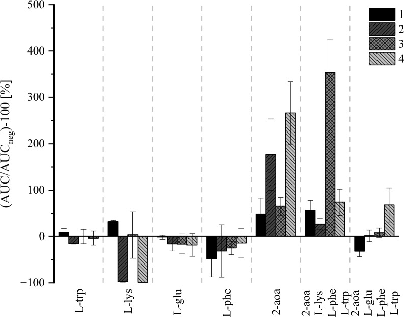
Fig. 4.
Relative formation of the four apicidins in medium supplemented with various amino acids (50 mM) in relation to non-supplemented medium. The Y-axis depicts the obtained area under the curves (AUC) after peak integration placed in relation to negative control (AUCneg) without supplementation in %. Black columns show apicidin F (1), dark grey columns show apicidin J (2), grey columns show apicidin K (3), and light grey column show the new apicidin L (4). Each experiment has N = 6 replicates cultivated for 5 d at 28 °C in the dark at saturated humidity, given are mean values ± standard deviations in %

