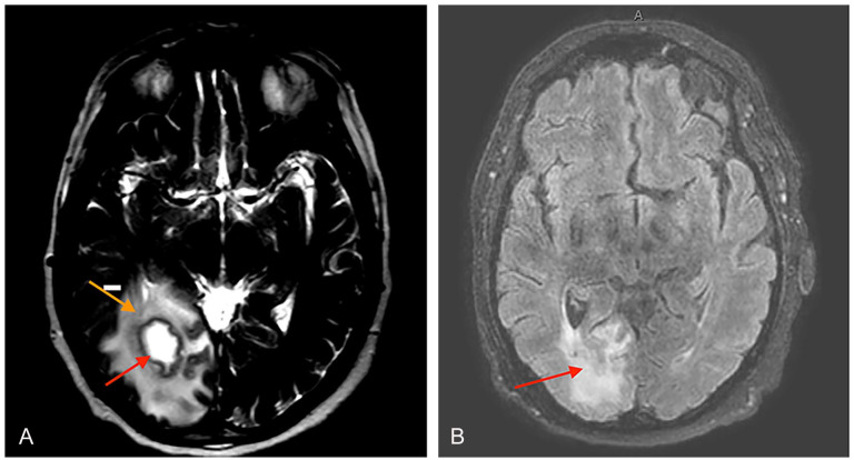
Figure 2.
(A) MRI brain with and without IV contrast showing a right parietal-occipatal lobe brain abscess (red arrow) with vasogenic edema (orange arrow). (B) MRI brain with and without IV contrast showing post-surgical changes and edema (red arrow) at the right parietal burr hole without evidence of residual right parietal-occipital brain abscess.

