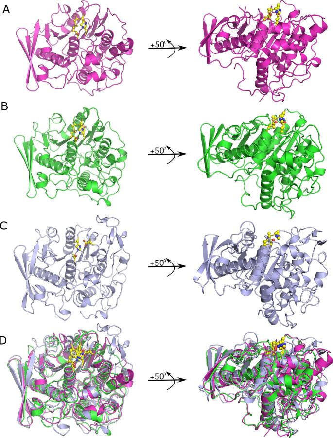
Fig. 2.
Comparison of the fold of rumen glucuronoyl esterases within the carbohydrate esterase family 15 esterase family. (A) Fibrobacter succinogenes (FsCE15), (B) Piromyces rhizinflata (PrCE15), and (C) Ruminococcus flavefaciens (RfCE15) adopt the canonical α/β-hydrolase fold that is seen in all of the GE structures solved to date. (D) Structure-based least squares structural superposition of FsCE15, PrCE15 and RfCE15. Catalytic residues, and the conserved disulfide bridge in the active sites of FsCE15 and PrCE15 are shown as yellow ball and sticks. Figures were generated with PyMol

