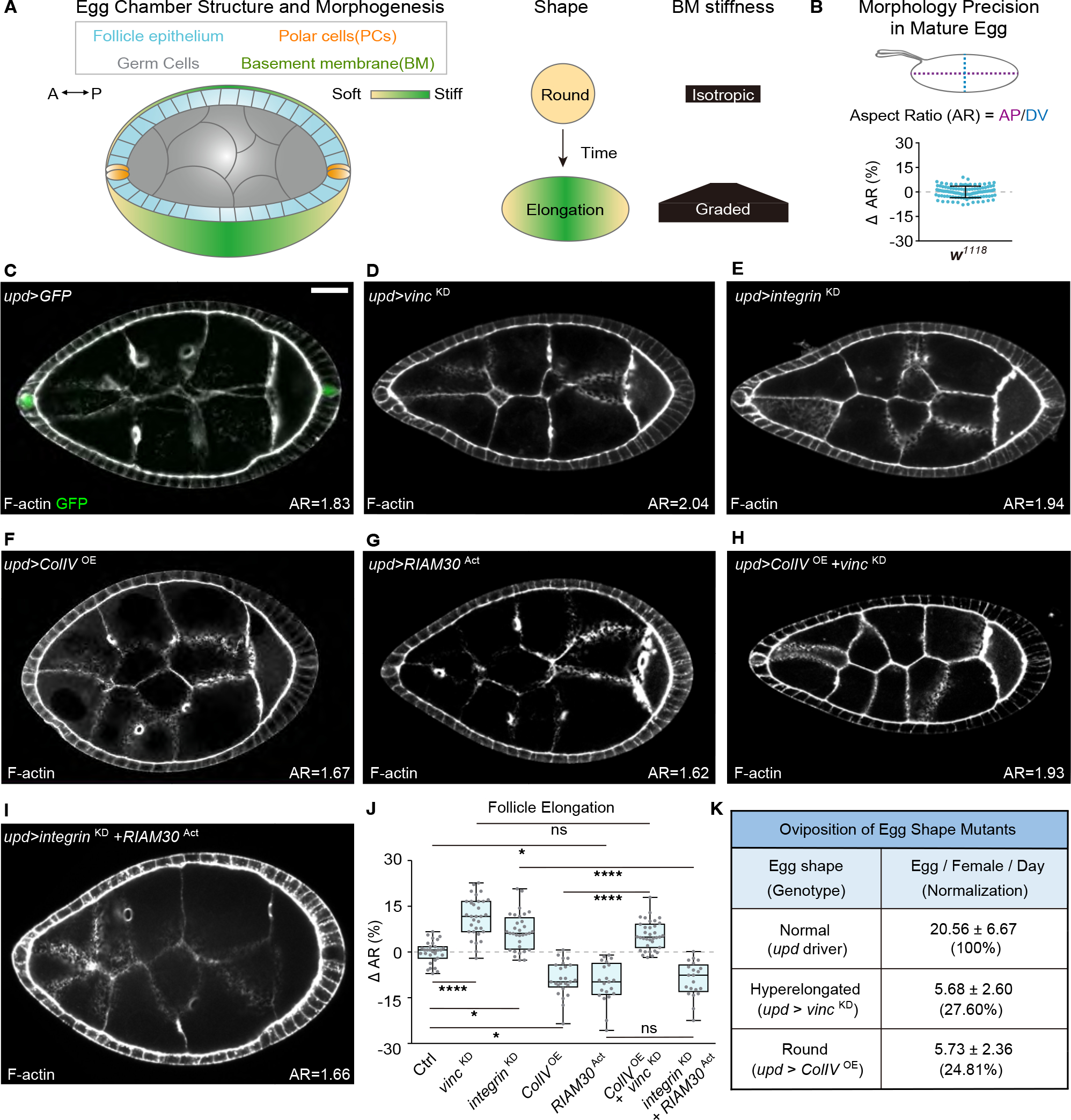
Figure 1. Polar cells direct tissue elongation through BM-responsive focal adhesion signaling.

(A) Overview of follicle elongation, showing cell types involved, along with mechanical patterning of the ECM that drives morphogenesis.
(B) Variation of aspect ratio (AR: AP/DV length) in wild-type eggs (w1118, n=108). Bar graph is mean ± standard deviation (0 ± 3.55%) subtracted to mean.
(C) Location of PCs, as displayed by upd-GAL4-driven GFP (n=19). F-actin marks follicle morphology. Mean AR is indicated in bottom right from this panel onward. Scale bars hereafter are 20 μm unless otherwise indicated.
(D-G) PC-specific knockdown (KD) of focal adhesion components Vinc (D, n=32) or Integrin (E, n=32) induces follicle hyperelongation, while overexpression (OE) of ColIV (F, n=25) or RIAM30Act (G, n=21) causes hypoelongation.
(H) Overexpression of ColIV with simultaneous depletion of Vinc (n=35) shows that Vinc acts downstream of ColIV in follicle shaping.
(I) Overexpression of RIAM30Act in PCs reverses the hyperelongation phenotype of Integrin depletion (n=32).
(J) Quantitation of follicle elongation in Ctrl (C, upd driver, n=31) and D-I. Statistics are shown in box and whiskers (Min to Max) plot, with comparisons performed using ANOVA with Dunnett’s multiple comparisons. *P < 0.05, **P < 0.01, ***P < 0.001, ****P < 0.0001, and n.s., not significant (P > 0.05).
(K) Oviposition rates for eggs with elongation phenotypes generated by PC manipulation. n for upd, upd>vinc KD, and upd>ColIV OE = 2056, 227, and 344 respectively.
