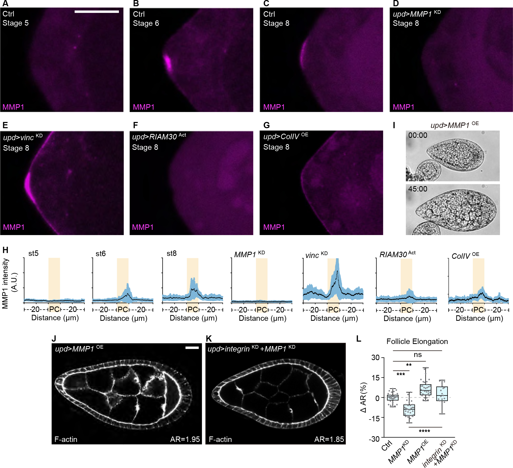
Figure 5. Focal adhesion-regulated MMP1 from PCs promotes tissue elongation.

(A-G) Images of anterior pole of developing follicles in ice preps. In control, MMP1 can be detected from stage 6 (A, stage 5, n=7; B, stage 6, n=15; C, stage 8, n=20). Staining of MMP1 is absent upon MMP1 depletion in the PCs (D, n=6). PC focal adhesion depletion leads to increased levels (E, n=13), while MMP1 in the PCs is reduced upon focal adhesion activation (F, n=9) or ColIV overexpression (G, n=16). Scale bars = 10 μm.
(H) Quantitation of MMP1 signal intensity along the follicle anterior pole, centered around the PCs (highlighted in yellow) for a total of 50 μm.
(I-J) Overexpressing MMP1 in PCs results in follicles that resist bursting upon osmotic shock (I, quantitation in Figure 3H–J, n=21) and is sufficient to drive hyperelongation (J).
(K) Co-depletion of MMP1 attenuates follicle hyperelongation induced by Integrin depletion from PCs.
(L) Quantitation of follicle elongation from Ctrl (upd driver, n=31), D (n=32), I (n=35), and J (n=19). Statistics used ANOVA with Dunnett’s multiple comparisons.
