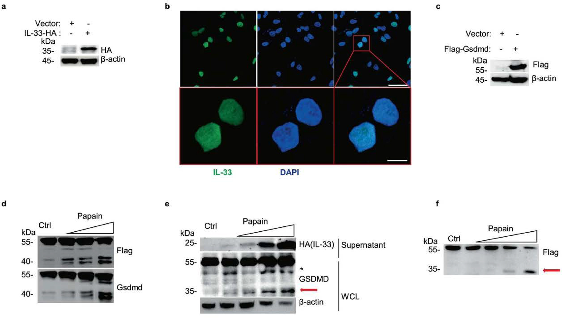
Extended Data Fig. 2 |. Construction of IL-33 and Gsdmd constitutively expressive airway epithelial cells.

a, Immunoblot analysis of whole-cell lysis (WCL) of constructed C-terminal HA-tagged IL-33 expressing A549 with Lenti-virus overexpression. b, Microscopy of the immunofluorescence stained IL-33 (green) and DAPI (blue) in A549-IL-33 cells without stimulations. Scale bars (50 μm upper, 10 μm below). c, Immunoblot analysis of constructed N-terminal Flag-tagged Gsdmd expressing MLE-12. d, Immunoblot analysis of MLE-12-flag-Gsdmd cells treated with papain (0 μg, 5 μg, 10 μg and 50 μg per well) for 30 min. e, Immunoblot analysis of WCL and culture supernatants of constructed IL-33 expressing A549 after treatment with papain (0 μg, 1 μg, 5 μg and 10 μg) for 30 min. f, Immunoblot analysis of WCL of N-terminal Flag-tagged GSDMD expressing A549 cells exposed to papain (0 μg, 5 μg, 10 μg and 50 μg) for 30 min. The red arrow marked fragment represents the activated form of p35 NT-GSDMD. Asterisk marked nonspecific fragment were not discussed in our work. Data are representative of two independent experiments with similar results.
