
Fig. 6 |. Gsdmd contributes to type 2 inflammatory immune responses.
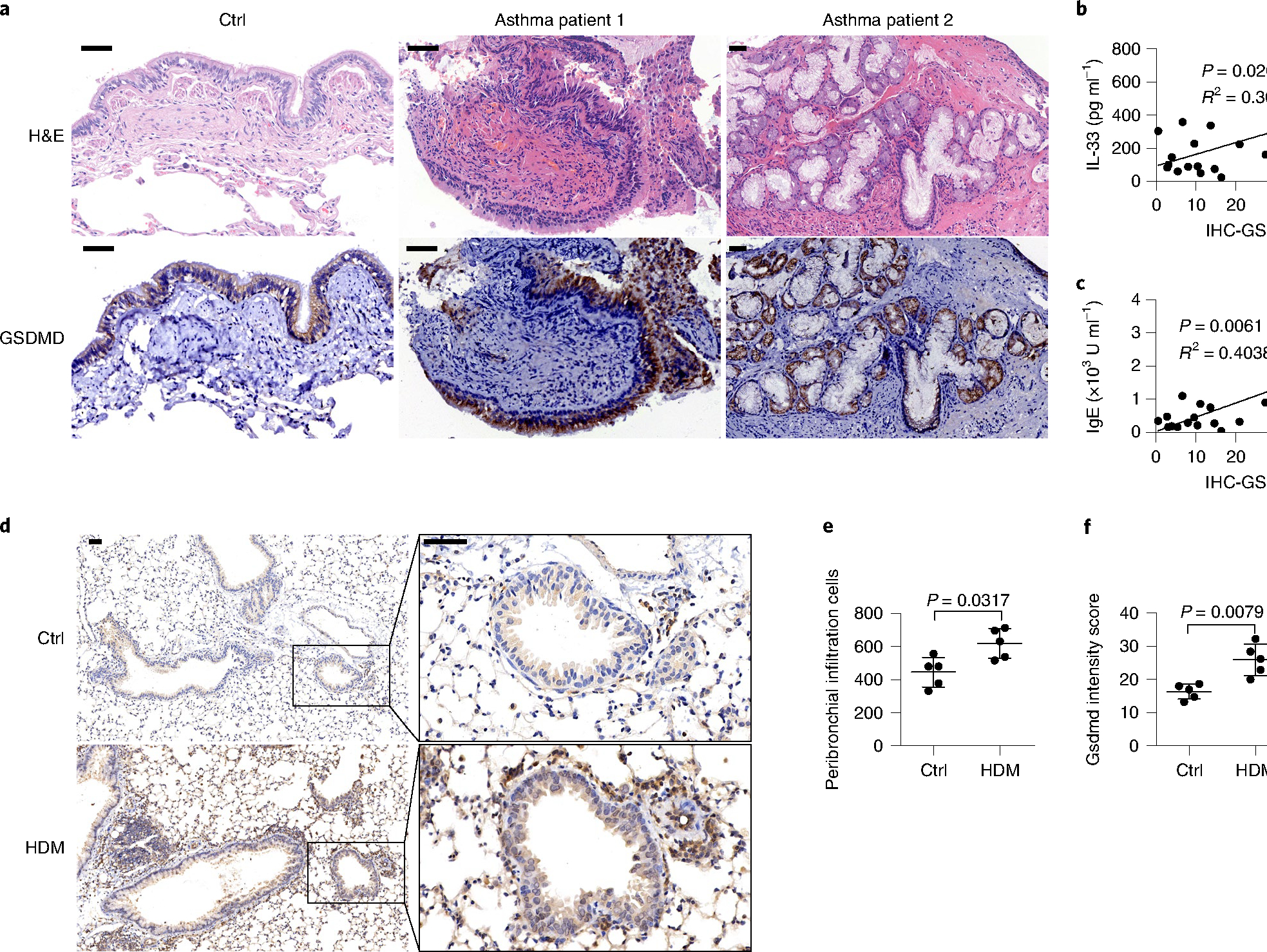
a, Microscopy images of H&E staining and immunohistochemical (IHC) staining of human GSDMD (brown) in bronchial tissue from people with and without asthma. b, IL-33 production in the BALF versus GSDMD expression in lung tissues of people with asthma. c, Serum IgE versus GSDMD expression in lung tissues of people with asthma. b,c, n = 17, two-tailed Pearson correlation. Linear regression curves fitted to these data are provided, alongside P values and R2 values. d, Microscopy imaging of immunohistochemically stained mouse Gsdmd protein in lung tissues. BALB/c mice were administered with HDM or PBS intranasally for up to 3 weeks to induce asthma symptoms. e, Histological evaluation of peribronchial inflammatory cell infiltration as in d. f, Statistical analysis of the Gsdmd protein intensity scores as in d. Error bar, mean ± s.e.m. e,f, n = 5, using the two-tailed Mann–Whitney U-test. Scale bars: a,d, 50 μm. a–f, Data are representative of at least three independent experiments.