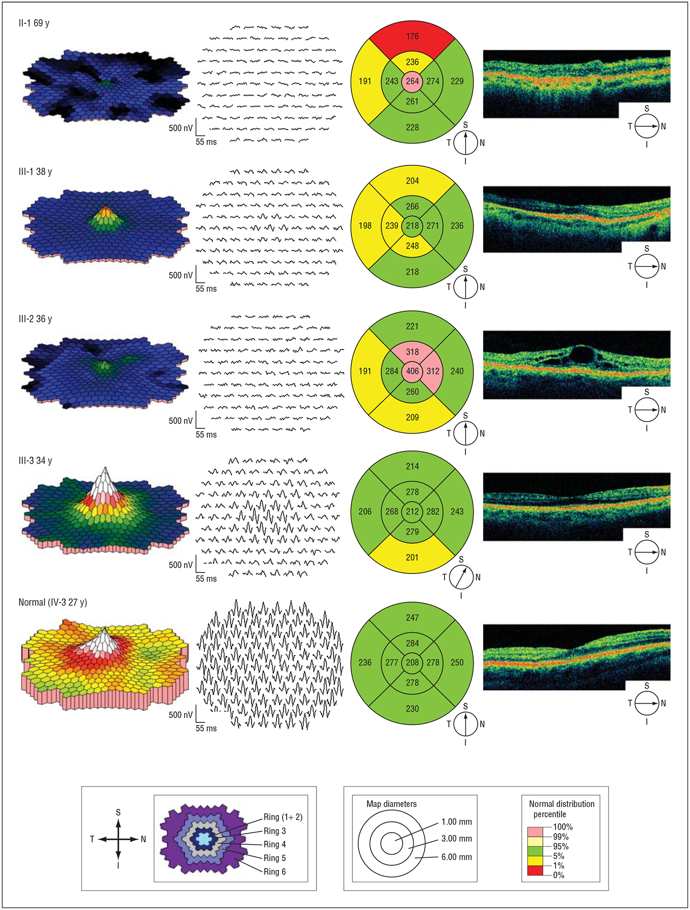
Figure 4.

Multifocal electroretinography and optical coherence tomography results of retinal function and morphologic structure in the macular region. Shown are 4 reexamined members from family 101 with KLHL7 mutation and 1 family member without the mutation. Multifocal electroretinography, trace arrays, and plots are retinal views from right eyes. Shown at the bottom left are multifocal electroretinography definitions of ring areas 1 through 6. Optical coherence tomography, line images, and macular thickness maps are from right eyes. Shown at the bottom right are optical coherence tomography definitions of macular thickness maps (in microns). I indicates inferior; N, nasal, S, superior; and T, temporal.
