
Fig. 3.
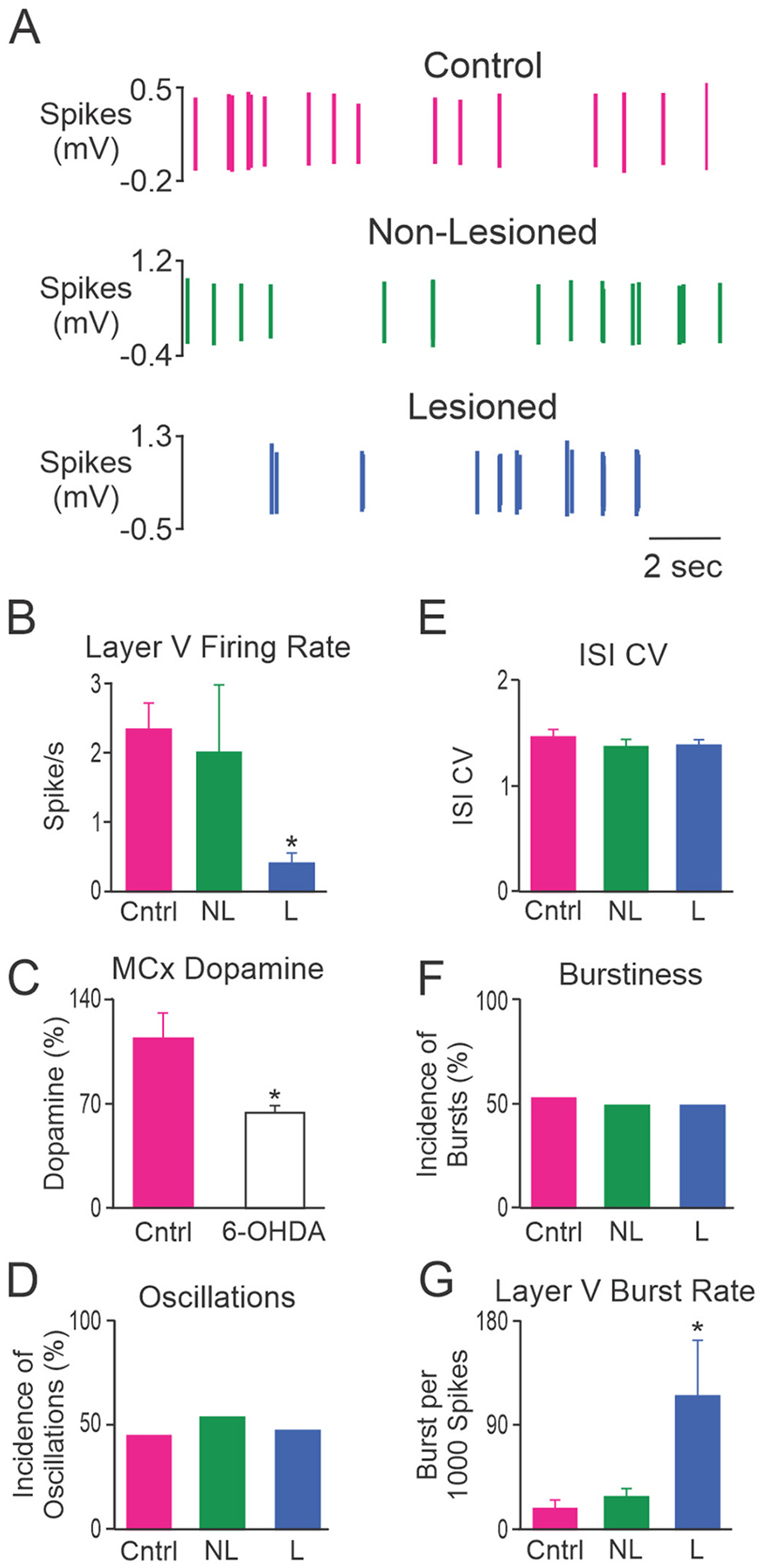
Characteristics of MCx spike trains from putative pyramidal neurons. A, Representative example of putative pyramidal neuron spike trains from control rats (Cntrl; top, red), and non-lesioned (NL; middle, green) and dopamine lesioned (L; bottom, blue) hemispheres of unilateral 6-OHDA lesioned rats. These 3 neurons were recorded in layer V and had firing rates of 2.84 spikes/s, 2.77 spikes/s and 0.19 spikes/s, respectively. Grouped data showing: B, the mean firing rate of putative pyramidal neurons in layer V; C, MCx dopamine content detected with HPLC and expressed as a percentage of the non-lesioned hemisphere; D, incidence of spike trains with significant 0.3–2.5 Hz oscillatory activity; E, mean ISI CV; F, incidence of bursty spike trains; and G, burst rate for putative pyramidal neurons in layer V. Layer V putative pyramidal neurons fired less frequently and with more bursts in the lesioned hemisphere relative to control rats. * Significant difference of p < 0.05 compared to control rats. Total cells: n = 56 from 9 Cntrl rats, n = 51 from 26 NL hemispheres and n = 75 from 30 L hemispheres. Layer V cells: n = 18 from 6 Cntrl rats, n = 17 from 9 NL hemispheres and n = 13 from 8 L hemispheres.