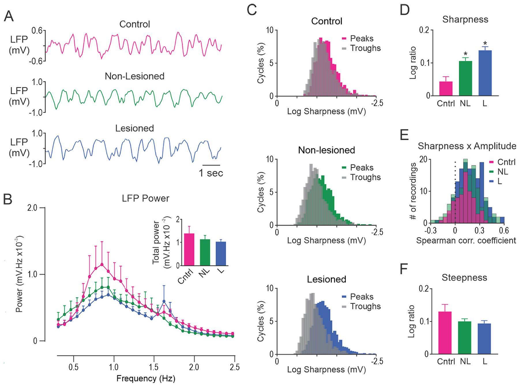
Fig. 4.

Unilateral dopamine lesion alters the shape of slow-wave oscillations in MCx but does not alter FFT power. A, Representative segments of MCx LFP activity in control rats (Cntrl; top, red), and non-lesioned (NL; middle, green) and dopamine lesioned (L; bottom, blue) hemispheres of unilateral 6-OHDA lesioned rats. B, FFT power spectral density over the 0.3–2.5 Hz range. Total power was not different between the groups (inset). C, Histograms showing the distribution of slow-wave peak and trough sharpness in a representative 300 s LFP recording in control rats (top), and non-lesioned (middle) and lesioned (bottom) hemispheres in unilateral 6-OHDA lesioned rats. D, Sharpness ratio was significantly larger in non-lesioned and lesioned hemispheres relative to control rats. E, On average, a positive correlation between cycle sharpness ratio and amplitude was observed for LFPs recorded from control rats and both hemispheres in 6-OHDA lesioned rats. F, Steepness ratio did not differ between control rats, non-lesioned and lesioned hemispheres. * Significant difference of p < 0.05 compared to control rats. LFP’s recorded from different electrode tracks were averaged to give a single data point for each rat. n = 14 Cntrl, n = 31 NL and n = 43 L.
