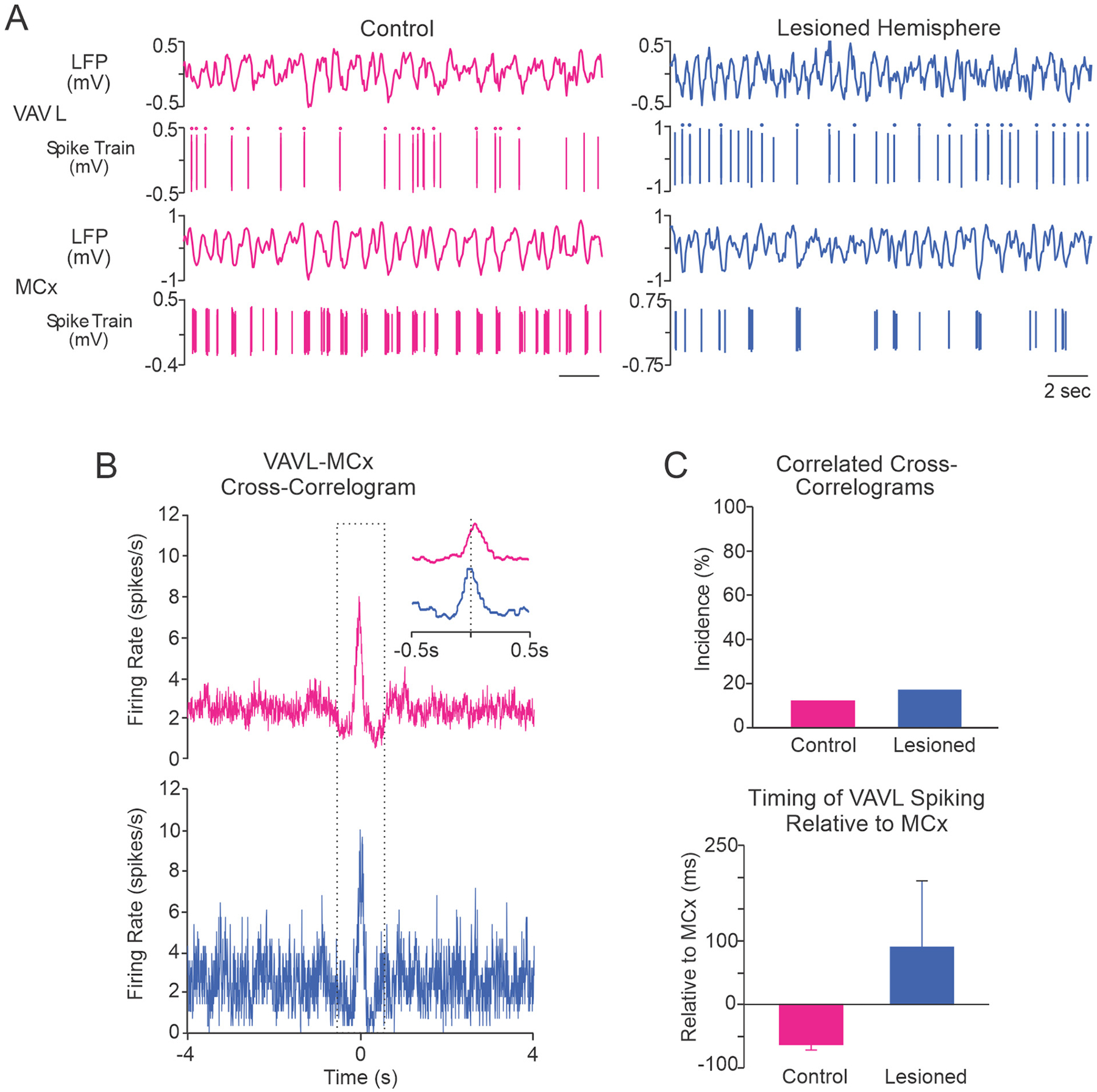
Fig. 5.

Correlated activity between VAVL neurons and putative pyramidal neurons in MCx. A, Representative examples of simultaneously recorded spike trains and LFPs from VAVL and putative pyramidal neurons in MCx show coincident oscillations in spiking activity in the control (red) and dopamine lesioned hemisphere (blue). The dots above spikes in spike trains indicate LTS bursts. B, MCx-triggered VAVL cross-correlograms from representative spike trains from control and dopamine lesioned hemispheres shown in A above. Cross-correlogram in the control hemisphere (top) shows that neuronal spiking was significantly correlated between VAVL and MCx. The peak in VAVL spiking occurred 46 ms before MCx spikes. For the lesioned hemisphere (bottom), the peak in VAVL spiking occurred 210 ms after MCx spikes. C, Population data of paired VAVL-MCx recordings. There was no significant difference between control rats (n = 25) and the lesioned hemisphere (n = 24) with respect to the proportions of VAVL-MCx spike train pairs with correlated activity (top) or the timing of VAVL firing relative to MCx (bottom).
