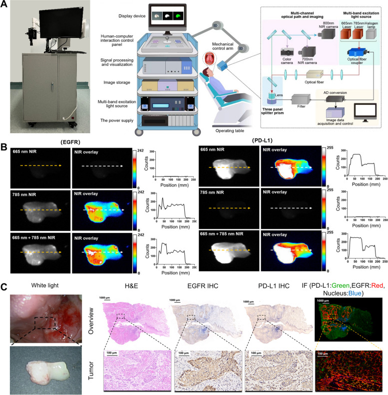
Fig. 5.
Nimotuzumab-ICG and Atezolizumab-Cy5.5 imaging probes were used to detect fresh OSCC biopsy samples. A The working model diagram and prototype of the home-made multispectral fluorescence imaging system. B Representative NIR and NIR overlay image of Nimotuzumab-ICG and Atezolizumab-Cy5.5 staining of fresh OSCC biospecimen from patients. Orange and white arrows correspond to the position and orientation of the cross-sectional fluorescence intensity profiles of the NIR and NIR overlay regions, respectively. Quantitative analysis showed that the fluorescence intensity in the tumor area and the para tumor area. NIR, Near-infrared. C White light, H&E, EGFR IHC, PD-L1 IHC, and IF staining of fresh biospecimen from OSCC patients. Scale bar, 1,000 μm and 100 μm

