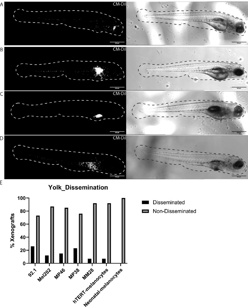
Figure 1.
Different inoculation sites in zebrafish UM xenografts. (A) Zebrafish xenograft injected behind the eye (retro-orbital) with 92.1 cells at 3 dpi. (B) Zebrafish xenograft injected in the perivitelline space with 92.1 cells at 3 dpi. (C) Zebrafish xenograft injected in the yolk sac with 92.1 cells that did not show cell dissemination at 3 dpi. (D) Zebrafish xenograft injected in yolk sac with 92.1 cells showing cell dissemination at 3 dpi. (E) Amount of dissemination present in xenografts injected in the yolk with 92.1 (n = 42), Mel202 (n = 31), MP46 (n = 40), MP38 (n = 34), MM28 (n = 28), hTERT-melanocytes (CRL-4059, n = 26), and neonatal melanocytes (GM21807, n = 35).

