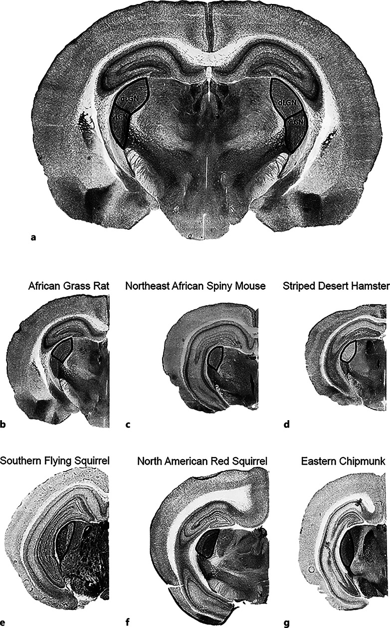
Fig. 3.
Photomicrographs of AChE-stained brain sections illustrating boundaries of dorsal lateral geniculate nucleus (dLGN) and ventral lateral geniculate nucleus (vLGN); the latter includes the intergeniculate leaflet (IGL). a African grass rat with dLGN and vLGN boundaries highlighted; African grass rat (b), Northeast African spiny mouse (c), striped desert hamster (d), southern flying squirrel (e), North American red squirrel (f), and eastern chipmunk (g) with dLGN boundary highlighted. Brains are not to scale. Boundaries identified using Paxinos and Watson [75].

