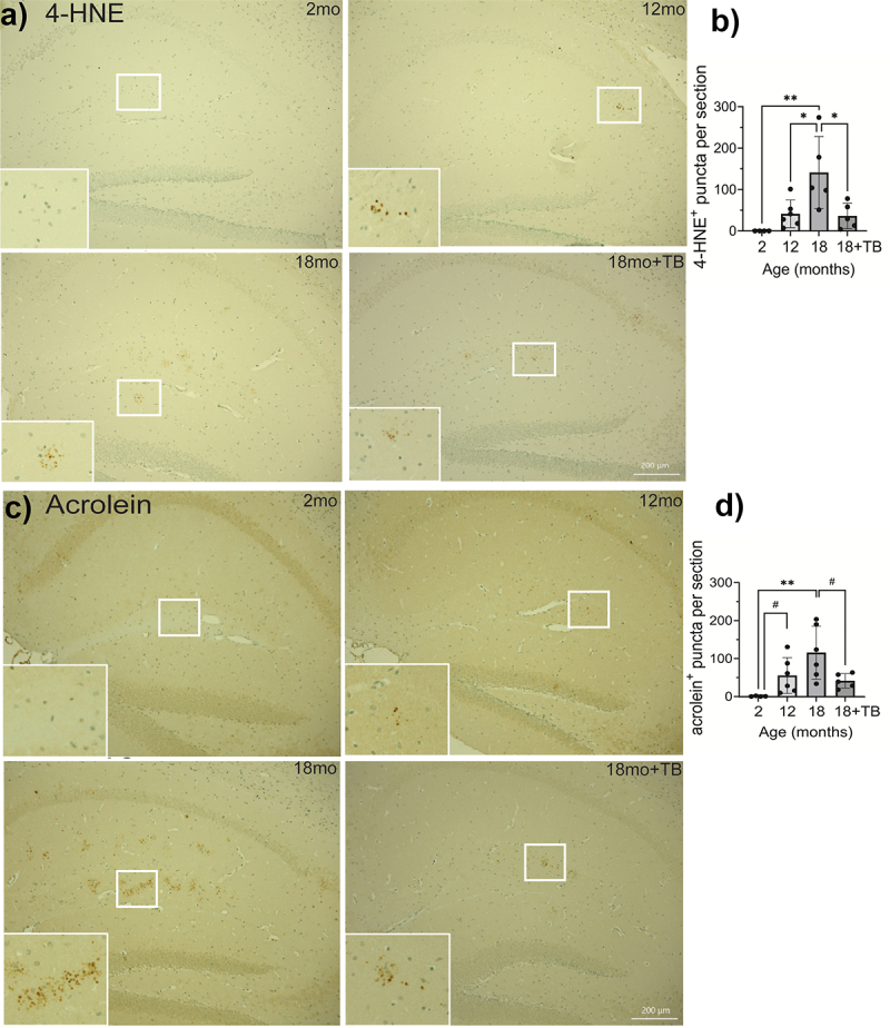
Figure 5.

Age-associated oxidative stress is abrogated in 3×Tg mice through oral TB administration.
Representative images of IHC staining for 4-HNE a) or acrolein adducts c) in sagittal brain sections in the hippocampus were visualized with DAB to assess oxidative stress. Quantification of individual puncta in 4-HNE b) or acrolein adducts d) stained sections was counted using Keyence Analyzer software as described in the Methods section. The majority of positive staining events was localized in the stratum lucidum and the molecular layer of the hippocampus. Magnification bars = 200 µm (a and c, 18mo+TB, white bars on bottom right). Bar graphs and error bars (b and d) represent mean scores ± SD, and data from individual mice are shown in closed circles. Statistics were calculated using parametric ordinary one-way ANOVA with multiple comparisons, and significance is indicated as *p < 0.05, **p < 0.01, or ***p < 0.001. Pairwise comparisons were calculated using unpaired t tests with the Welch’s correction, which does not assume equal SD, with significance indicated as #p<0.05.
