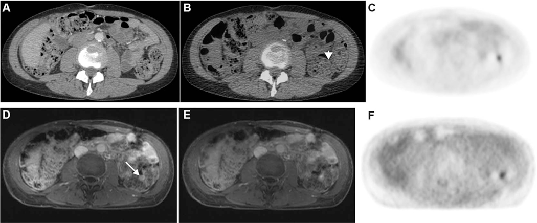
FIGURE 1.
Top panel: CE-CT (A), fused PET/CT (B), and FDG-PET images (C). Bottom panel: CE-MRI (A), fused PET/MRI (B), and FDG-PET images (C). A51-year-old female with a history of high-grade serous ovarian cancer status post complete cytoreduction. A small focus of moderate uptake along the wall of the descending colon (arrowhead in B) does not have an abnormal anatomic correlate on CE-CT to suggest metastasis and was attributed to physiologic bowel uptake. However, on PET/MRI, the focal uptake corresponds to an enhancing nodule on the surface of the colon (arrow in D). This lesion was consistent with a serosal metastasis and was subsequently resected.

