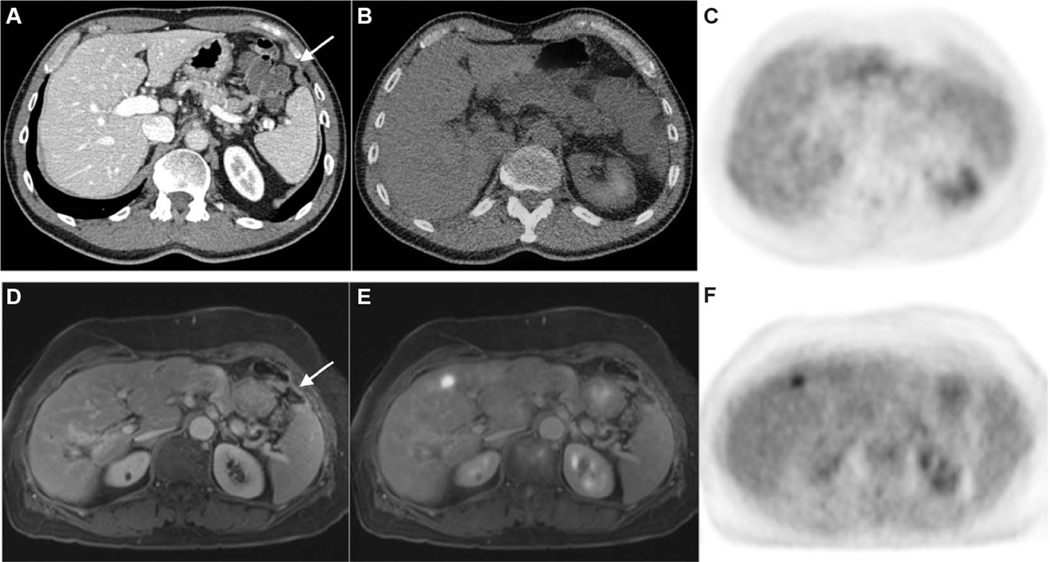
FIGURE 2.
Top panel: CE-CT (A), fused PET/CT (B), and FDG-PET images (C). Bottom panel: CE-MRI (D), fused PET/MRI (E), and FDG-PET images (F). A 65-year-old male with a history of resected adenocarcinoma of the ascending colon. A thin rind of soft tissue (arrow in A) is difficult to discern from the diaphragm on CT, and the degree of radiotracer uptake is similar to hepatic parenchyma on PET and fused PET/CT images. An area of abnormal peritoneal enhancement in the left upper quadrant (arrow in D) is readily perceptible on CE-MRI. This was confirmed to be metastatic adenocarcinoma following surgery. In this case, PET/MRI was able to detect peritoneal metastases approximately 4 months before a positive SCI scan that led to surgery, at which point widespread unresectable disease was found intraoperatively.

