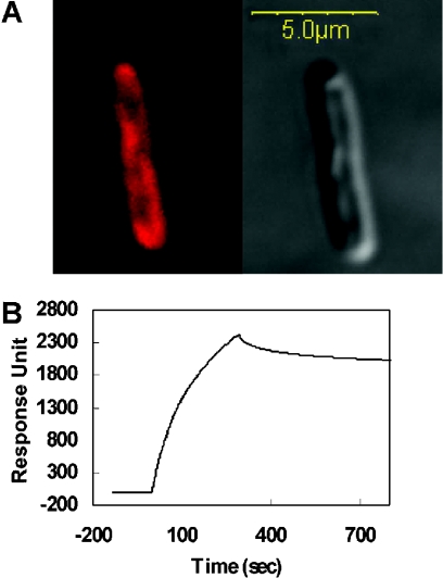
Figure 7. Localization of the Tar-1 peptides to the bacterial membrane.
(A) Partitioning of rhodamine-labelled Tar-1 R/R to the inner membrane of E. coli was determined by confocal laser-scanning microscopy. A similar result was observed for the Tar-1 WT and Tar-1 E/E peptides. The Tar-1 WT peptide was previously shown to be localized to the bacterial membrane. The fluorescence image is on the upper-left-hand side and the transmission light image is on the upper-right-hand side. Most of the peptide is localized on the plasma membrane, although some peptide also penetrates into the cytoplasm. Confocal images were obtained using an Olympus IX70 FV500 confocal laser-scanning microscope. There were only minute background fluorescence intensities observed in the control bacteria. (B) A sensogram of the binding between Tar-1 WT peptide (10 μM) and PE/PG (7:3, w/w) bilayers.

