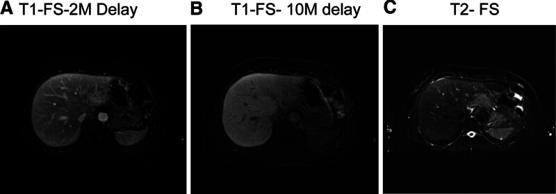
Figure 3.
MRI performed 1 mo after the completion of radiation therapy. MRI revealed an irregularly shaped mass in the left lateral segment of the liver exhibiting high signal intensity on T2-weighted images and subtle enhancement. These imaging features suggested the involvement of lymphoma in the left lateral hepatic segment. FS = fat suppressed imaging, M = minutes, MRI = magnetic resonance imaging.

