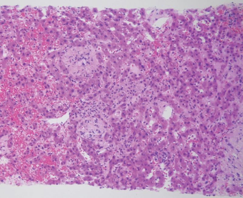
Figure 4.
Histologic features of liver biopsies. Hematoxylin and Eosin (HE) staining, original magnification: 200×. Histopathological examination of the liver biopsy specimen revealed sinusoidal denudation, sinusoidal dilatation, and hemorrhage. Additionally, there was evidence of zone 3 (centrilobular) fibrosis and occlusion of the central vein. The liver tissue exhibited a characteristic sinusoidal and endothelial cell injury pattern.

