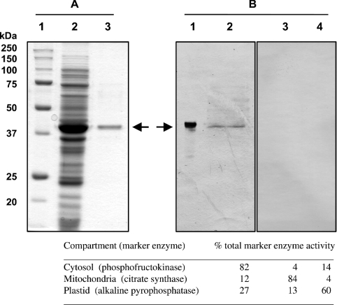
Figure 2. SDS/PAGE and immunoblot analysis of Arabidopsis NADHK.
(A) SDS/PAGE [12% (w/v) resolving gel] of recombinant NADHK purified from E. coli. Lane 1 contains 10 μg of various protein standards. Lane 2 contains 30 μg of a crude E. coli protein extract prepared after 3 h induction with 500 μM isopropyl β-D-thiogalactopyranoside. Lane 3 contains 2 μg of NADHK purified on S-protein–agarose. Protein staining was performed with Coomassie Blue R-250. (B) Immunoblot analysis performed using anti-NADHK serum. Lane 1 contains 0.05 μg of recombinant NADHK purified on S-protein–agarose. Lanes 2–4 each contain 50 μg of protein extracted from 7-day-old Arabidopsis cell suspension cultures enriched for various cellular compartments as follows (marker enzyme given in parentheses): lane 2, cytosol (phosphofructokinase); lane 3, mitochondria (citrate synthase); lane 4, plastid (alkaline pyrophosphatase). Antigenic polypeptides were visualized using alkaline phosphatase-tagged secondary antibody.

